Cerebral Perfusion and Autoregulation
Cerebral perfusion depends on the interaction among large-vessel hemodynamics, microvascular resistance, tissue demand, venous drainage, and active vascular regulation. We develop patient-specific multiscale models that connect image-derived vascular networks with three-dimensional tissue-perfusion domains. These models incorporate physiologic supply–demand relationships, passive vessel mechanics, and arteriolar autoregulation to quantify how systemic pressure, posture, vascular stiffness, and arterial stenosis affect regional cerebral blood flow.
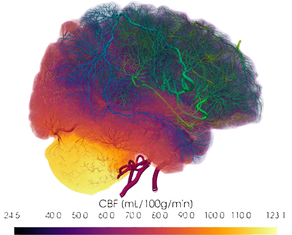
Physiologic Flow Distribution and Perfusion Territories
Realistic cerebrovascular simulations require boundary conditions that represent the blood-flow demand of the supplied brain tissue. We estimate subject-specific flow distributions by associating image-derived cerebral arteries with surrounding tissue regions. The required flow for each territory is determined from tissue mass and regional cerebral blood-flow demand and is distributed through a one-dimensional vascular model.
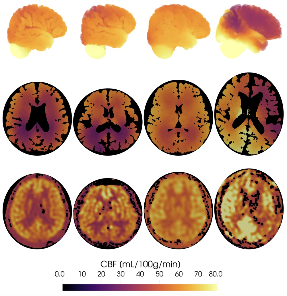
The model is evaluated by comparing estimated arterial flow distributions and perfusion territories with reported physiological data and perfusion imaging. The supply–demand formulation also reduces sensitivity to variations in segmented vessel caliber compared with geometry-only flow allocation.
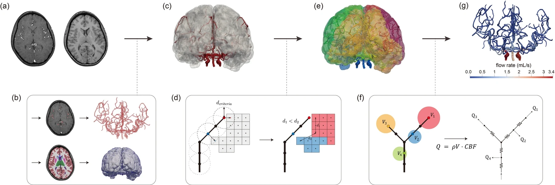
Multiscale Cerebral Perfusion Modeling
Cerebral vessels below the resolution of clinical imaging cannot be reconstructed directly. We extend image-derived arteries with synthetic vascular trees and couple one-dimensional blood-flow equations with a three-dimensional tissue-perfusion model. This multiscale formulation transfers pressure and flow between the explicit vascular network and the tissue domain and provides spatial estimates of cerebral perfusion.
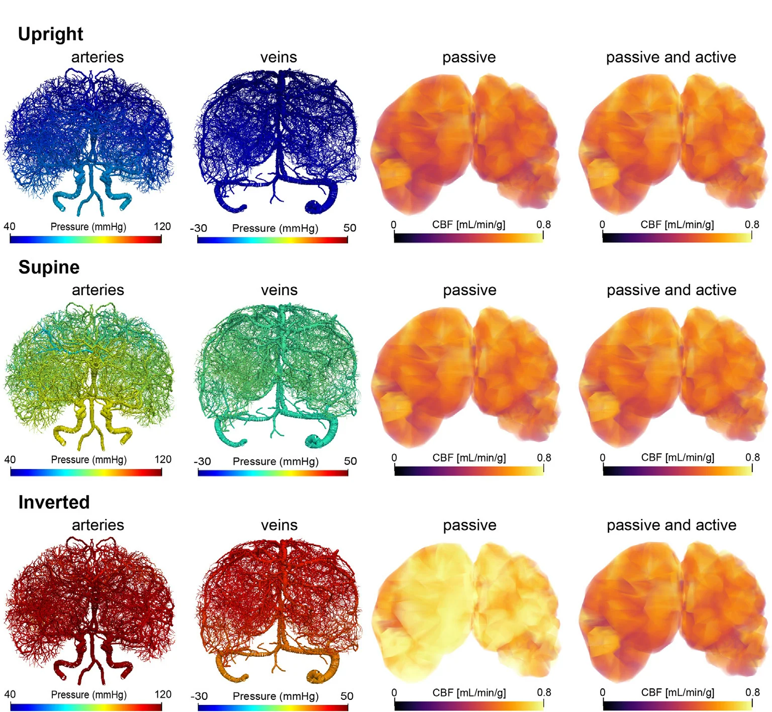
Autoregulatory Coupling under Pressure and Postural Changes
Cerebral autoregulation modifies arteriolar resistance to stabilize blood flow under changes in systemic and local conditions. We incorporate active arteriolar dilation and constriction into a multiscale model that includes arterial and venous networks, pre- and postcapillary compartments, and a three-dimensional perfusion domain. This framework allows the coupled effects of aortic pressure, gravity, vessel-wall stiffness, and metabolic demand to be examined.
Under the investigated conditions, active autoregulation maintains cerebral blood flow within a relatively narrow range by adjusting arteriolar diameter. Arterial deformation and total vascular volume are affected by systemic pressure and gravity, whereas venous volume is primarily sensitive to posture. Increased vascular compliance amplifies posture-dependent volume changes and the corresponding autoregulatory response.
Autoregulatory Response to Ischemia
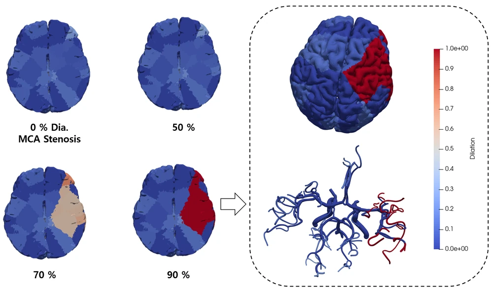
Arterial stenosis reduces downstream pressure and produces a mismatch between tissue supply and metabolic demand. We model arteriolar vasodilation as a reduction in downstream vascular resistance in response to local hemodynamic and metabolic stimuli. This mechanism allows the contribution of active regulation to perfusion recovery to be quantified under controlled stenosis conditions.
Under the investigated stenosis conditions, arteriolar vasodilation produces a larger improvement in tissue perfusion than sub-resolution collateral vessels alone. The contribution of collateral circulation remains dependent on vessel caliber, density, and stenosis severity, and is treated in Cerebrovascular Networks and Collateral Circulation.
Model Evaluation and Interpretation
We evaluate cerebral perfusion models through comparisons with physiological flow ranges, vascular morphology data, perfusion territories, and medical perfusion imaging. These comparisons assess whether the models reproduce plausible pressure, flow, and tissue-perfusion patterns. The resulting simulations provide mechanistic estimates rather than direct clinical diagnoses and remain dependent on assumptions regarding sub-resolution vasculature, tissue properties, and autoregulatory capacity.
Selected work
Multiscale modeling of posture-dependent cerebrovascular hemodynamics with autoregulatory coupling
Computers in Biology and Medicine 203:111502
Annals of Biomedical Engineering
Physiologic model of the cerebrovascular system using supply and demand between arteries and tissues
Scientific Reports 15:27785
Computer Methods and Programs in Biomedicine 244:107956